
Tuesday, February 12, 2013
Wednesday, January 23, 2013
Immunofluorescence confocal microscopy

Thursday, March 29, 2012
Immunofluorescence light microscopy
Dr. Bergstrom uses immunofluoresence to identify cells expressing both pMAPK and CAMKII.
Red = pMAPK green=CAMKII

Amygdala, 10x
Monday, February 13, 2012
pMAPK labeled principal neuron in the amygdala

pMAPK immuno-labeled neuron in the Lateral Amygdala - 100x objective lens, N.A. 1.4, oil immersion lens, light microscopy - Bregma -3.24
Wednesday, February 1, 2012
Lab members

LTC Jen Coyner is using the lab microscope and MBF Neurolucida program to identify pMAPK neurons in the mouse amygdala.
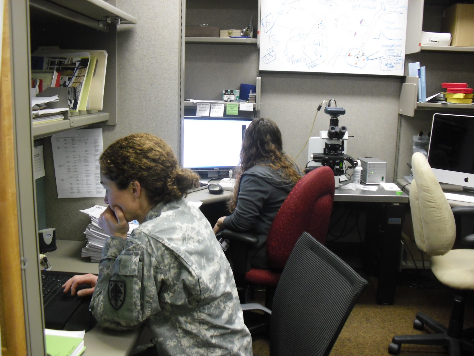
Anna learning how to identify neurons and Jen concentrating hard on her next move on her mouse project.

Dr Bergstrom
Saturday, January 21, 2012
SFN Snapshot

Dr. Bergstrom in action at SFN 2011 in DC. He drew quite a crowd during his poster session. Here's why.
Friday, January 20, 2012
Dr. McGuire's SFN 2011 Poster
Check out Dr. Jennifer McGuire's Traumatic Brain Injury Poster presented at the 2011 SFN Conference in Washington DC.

To zoom in - Click on the picture, right click on the image that pops up. Select Open Image in New Tab.
Subscribe to:
Posts (Atom)